Micropattern-controlled chirality of focal adhesions regulates the cytoskeletal arrangement and gene transfection of mesenchymal stem cells (Wang et al., 2021)1
Chirality is a lack of mirror symmetry. Chiral molecules are a fundamental idea in organic chemistry, and organisms are obviously chiral – your heart is on the left, not the right, after all. In fact, the internal organs of all vertebrates are asymmetrically organised.2 What had never occurred to me before I saw this paper, though, is the idea of chirality at the cellular scale.
It’s not obvious why macroscopic asymmetry is advantageous, though it has been proposed that chirality in the heart improves efficiency of circulation3, so improved fluid dynamics or spatial packing seem plausible in principle. Even less clear is why macro-scale asymmetry is consistent across populations. In humans, around 1 person in 10,000 is born with situs inversus: all of their internal organs are reflected through the sagittal plane. Many of those people have no symptoms at all!
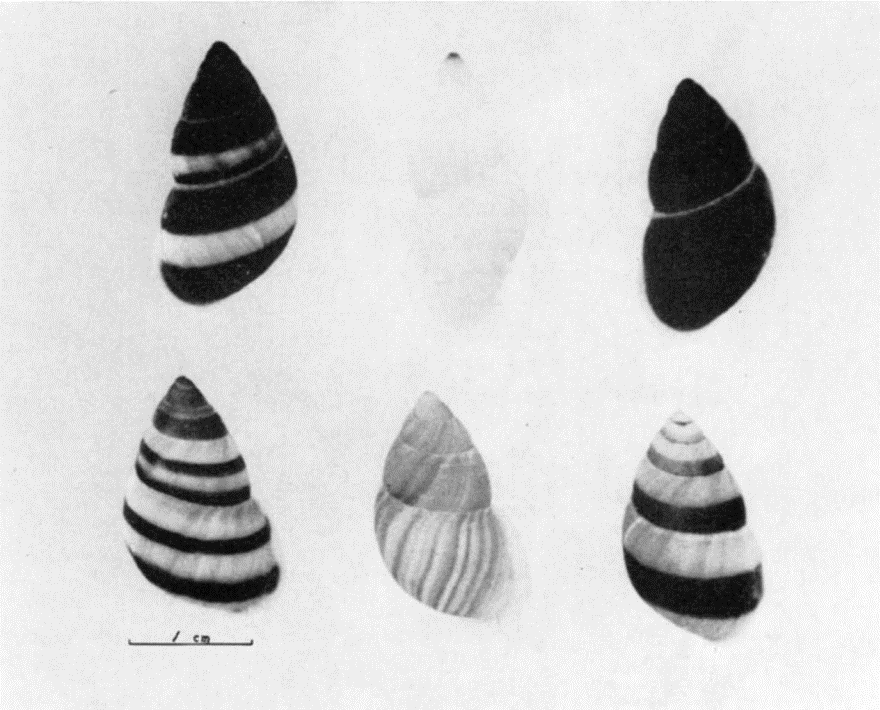
Another lovely example of this mystery is in snails with coiled shells (figure 1). These coils can be either left-handed or right-handed helices, and within each snail species the handedness of coiling is consistent. However, both left-hand and right-handed spiralling species exit.
 |
|---|
| Figure 1: Asymmetric Coiling of Snail Shells, reproduced from Murray and Clarke, 19664 |
The mechanisms by which symmetry is first broken in development are unknown. Left-right axes in molecular expression emerge early in embryonic development (as expected, given the heart is the first organ to form), but how asymmetric transcription first arises is an open question.
At the individual cell level, chirality has been observed in organelle distribution, in migration preferences, and in rotation of the nuclei.5,6 Interestingly, it was proposed that the nuclear rotation effects are driven by asymmetry in the actin cytoskeleton, which also clearly influences organelle distribution.7 Surely the cell skeleton is essential to this story?
What’s this paper trying to do then?
Given the importance of the actin skeleton to cell chirality noted above, focal adhesions which link that skeleton to the extracellular environment are a natural candidate for transmission of chiral information. Wang et al.1 generated surfaces with chiral patterns of fibronectin and used them to probe induction of chirality in adhered cells.
How did they do it?
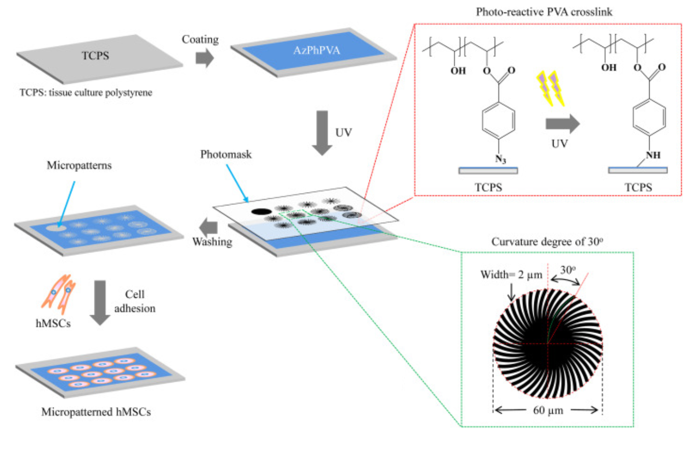
Spiral patterns of PVA were generated on a polystyrene surface by photolithography – the PVA was cross-linked to the polystyrene via UV-driven reactive of an azide linker under a photomask (figure 2). Both the degree and the angle of spiral were varied.
 |
|---|
| Figure 2: Generation of chiral fibronectin micropatterns, reproduced from Wang et al., 20211 |
What did they find?
Sadly, not much! While the angle of curvature of the spiral does have an impact - a 60° swirl induced shorter focal adhesions with less well-defined actin attachment, lower cellular stiffness (measured by AFM), and less efficient uptake of transfection vectors – the handedness of the spiral had no effect on any of their assays. To my mind that’s not a big shock: the fibronectin they’re using is of the same chirality either way, and the chirality of the spiral is too large-scale to be detected by an individual focal adhesion, which are typically on the order of ~2-3µm in length.8 It also makes more sense for chiral induction to be a function of molecular chirality than microscale surface chirality, given that development takes place in an environment where chiral molecules are everywhere – every protein or sugar present! – and one assumes the uterine lining is not covered in spiral micropatterns.
References
- Wang, Y. et al. Micropattern-controlled chirality of focal adhesions regulates the cytoskeletal arrangement and gene transfection of mesenchymal stem cells. Biomaterials 120751 (2021) doi:10.1016/j.biomaterials.2021.120751.
- Boorman, C. J. & Shimeld, S. M. The evolution of left–right asymmetry in chordates. BioEssays 24, 1004–1011 (2002).
- Kilner, P. J. et al. Asymmetric redirection of flow through the heart. Nature 404, 759–761 (2000).
- Murray, J. & Clarke, B. The Inheritance of Polymorphic Shell Characters in Partula (Gastropoda). Genetics 54, 1261–1277 (1966).
- Wan, L. Q., Chin, A. S., Worley, K. E. & Ray, P. Cell chirality: emergence of asymmetry from cell culture. Philos Trans R Soc Lond B Biol Sci 371, (2016).
- Chin, A. S. et al. Epithelial Cell Chirality Revealed by Three-Dimensional Spontaneous Rotation. PNAS 115, 12188–12193 (2018).
- Yamanaka, H. & Kondo, S. Rotating pigment cells exhibit an intrinsic chirality. Genes to Cells 20, 29–35 (2015).
- Kim, D.-H. & Wirtz, D. Focal adhesion size uniquely predicts cell migration. The FASEB Journal 27, 1351–1361 (2013).